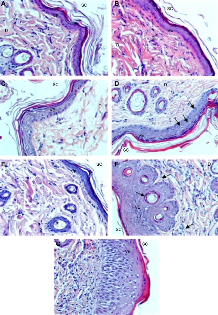
Figure 3.
Representative images of the skin sections.
Notes: (A) Control rats, skin treated with 100 μL of 0.8% formalin (B) and various concentrations of PAMAM-NH2 generation 2: (C) 3 mg/mL, (D) 6 mg/mL, (E) 30 mg/mL; (F and G) 300 mg/mL. Reported vacuolization of keratinocytes in the basal and spinous layers (arrows) (D), inflammatory infiltrations of leukocytes and connective tissue fibers hyperplasia (dermis) (F and G) and apoptotic keratinocytes (epidermis) and granulocyte infiltration in the upper dermis (F) (arrows). H and E staining, magnification ×200.
Abbreviations: PAMAM, polyamidoamine; SC, stratum corneum; VE, viable epidermis; D, dermis; H and E, hematoxylin-eosin.

