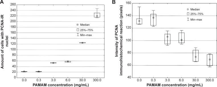
Figure 5.

Quantitative image analysis of PCNA-staining.
Notes: (A) Amount of cells with PCNA-IR nuclei, (B) intensity of immunohistochemical reaction in the rat skin sections treated with 100 μL of various concentrations of PAMAM-NH2 generation 2: 0.3 mg/mL, 3 mg/mL, 6 mg/mL, 30 mg/mL, 300 mg/mL (n=6).
Abbreviations: PAMAM, polyamidoamine; PCNA-IR, PCNA immunoreactivity; Min, minimum; Max, maximum.
