
Figure 2.
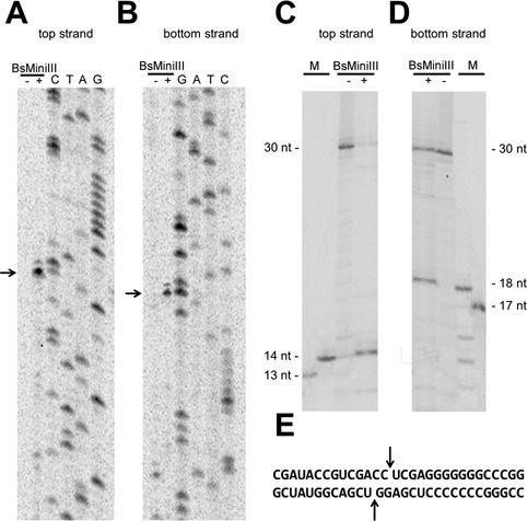
Identification of the BsMiniIII cleavage site in dsRNA. (A, B) Mapping of the cleavage site in the pKS-ACCU molecule by primer extension. The detection of the cleavage by primer extension reaction using a 33P-labeled primer complementary to the top strand (A) and to the bottom strand (B). The reactions were resolved on a denaturing polyacrylamide gel and visualized using autoradiography. The arrows indicate the strong termination of the primer extension. A dash and a plus sign indicate a primer extension reaction carried out on an uncleaved and a cleaved template, respectively. (C, D) Mapping of the cleavage site in a 30 bp dsRNA oligonucleotide (sequence shown in panel (E)). Detection of the cleavage of a 33P-labeled 30 bp fragment that corresponds to the top strand (C) and to the bottom strand (D) of the pKS-ACCU dsRNA molecule. A dash and a plus sign indicate an uncleaved and cleaved oligonucleotide, respectively. The 13, 14, 17 and 18-nt markers are 33P-labeled fragments of the 30-nt oligonucleotide shortened at the 3′ end. (E) The sequence of the dsRNA oligonucleotide substrate. The arrows indicate the cleavage site.