
Figure 3.
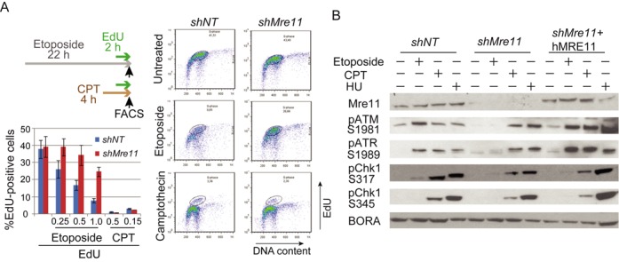
(A) Activation of intra-S and G2/M checkpoint in response to DNA damaging agents. Checkpoint activation was compromised in response to etoposide, but not to the replication inhibitor CPT in Mre11-KD cells. Experimental designs (top left): cells were treated either etoposide (22 h) or CPT (4 h). EdU was added for 2 h before FACS analyses. Right: profiles of cells with EdU incorporation for untreated cells (top), cells treated with etoposide (middle) and cells treated with CPT (bottom). Circles indicate cells with replicating DNA (labeled with EdU). Left bottom: histograms of EdU-positive cells. Several concentrations of agents were used. Note that EdU-positive cells are more abundant in Mre11-KD cells than in control cells when treated with etoposide, but not with CPT. (B) Activation of intra-S and G2/M checkpoint kinases in response to DNA damaging agents. Mre11-KD cells lack activation of ATM, ATR and Chk1 checkpoint kinases in response to etoposide, but not to replication inhibitors. Cells were treated as indicated, and activating phosphorylation of kinases was monitored by western blotting. Staining for Bora was used as a loading control.