
Figure 1.
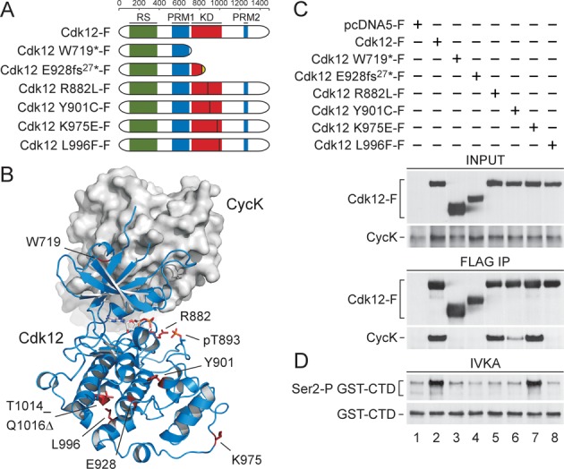
CDK12 mutations in HGS-OvCa abrogate the activity of Cdk12 predominantly by impairing the interaction between Cdk12 and CycK. (A) Schematic depiction of the wild-type and mutant Cdk12 proteins containing individual CDK12 mutations analyzed in this study. Highly structured kinase domain (KD; red), arginine/serine rich region (RS; green) and two regions with proline-rich motifs (PRM1 and PRM2; blue) are depicted. The ruler on top indicates the length of Cdk12 protein in amino acids. Vertical lines denote sites of individual missense and insertion mutations. Finally, ‘fs’ stands for a frame-shift mutation and the associated number indicates the number of altered amino acids at the C-terminus of mutant Cdk12 protein (dotted). (B) Overall structure of human Cdk12/CycK and positions of the mutations. Cdk12 is shown as cartoon representation in blue and CycK as surface representation in grey. CDK12 missense, insertion and internal deletion mutations are located in the C-terminal lobe of the KD. The mutated amino acid residues are highlighted in red. pT893 is highlighted in orange. (C) Effects of the CDK12 mutations on the interaction between Cdk12 and CycK. The indicated wild-type and mutant FLAG epitope-tagged Cdk12 proteins (Cdk12-F) were immuno-purified from whole cell extracts (WCEs) of the individual HEK 293 Flp-In T-Rex cell lines using FLAG-M2 agarose (FLAG IP) and examined for their interaction with endogenous CycK. Levels of Cdk12-F and CycK proteins in WCEs (INPUT, 5% of WCEs; top) and IPs (FLAG IP; bottom) were detected by Western blotting using FLAG and CycK antibodies. (D) CDK12 mutations abrogate the kinase activity of Cdk12. The indicated wild-type and mutant Cdk12-F proteins were immuno-purified (IP) as in panel C and the complexes were examined for their kinase activity by in vitro kinase assay (IVKA) toward the recombinant GST-CTD. Levels of Ser2-P GST-CTD isoforms and input GST-CTD (30%) were detected by Western blotting using Ser2-P-specific RNAPII and GST antibodies.