
Figure 1.
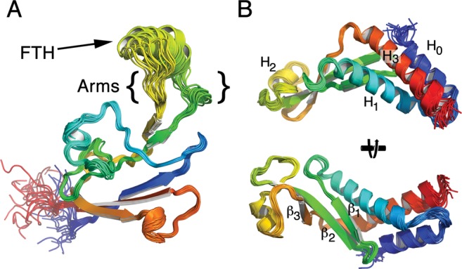
Structures of B. subtilis β-flap and NusAN. (A) Structure of β-flap shown as a chainbow from blue N-terminus to red C-terminus. FTH, β-flap tip helix. Arms, flexible regions lining the body of the β-flap to the FTH (see text for details). (B) NusAN shown as a chainbow from blue N-terminus to red C-terminus in two orientations. Bottom view is a rotation of the top view 90° into the page. The α-helices and β-strands are numbered in NusAN for reference in text.