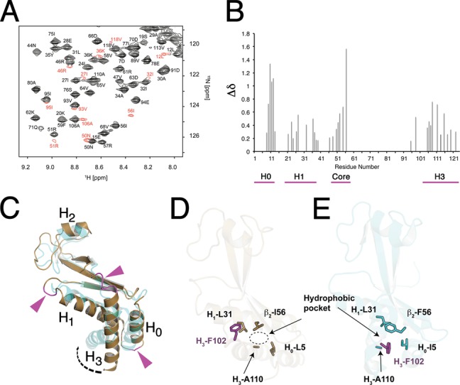
Figure 4.

NusAN is in equilibrium between a major and minor structural form. (A) A section of the HSQC spectrum of NusAN is shown with residues that exhibit a large difference in chemical shift in the minor conformation coloured in red. Note the particularly large chemical shift for I56 in the major (magenta asterisk) and minor (cyan asterisk) conformations. (B) Δδ plots based on chemical shift changes highlighting residues present in multiple conformations in B. subtilis NusAN plotted against residue number. Magenta line below indicates residues in the core and helical bundle in multiple conformations. (C) Superposition of the structure of B. subtilis (bronze) and E. coli (PDB 2KWP, cyan) NusAN showing how alternative conformations could form due to flexibility in regions of signal broadening (magenta sections and arrows) connecting the α-helices to the rest of the molecule. α-Helices are labelled from H0 to H3. (D, E) Identification of a hydrophobic pocket and rotation of F102 (magenta) around residue 56 in B. subtilis ((D); bronze) and E. coli ((E); cyan). NusAN structures.
