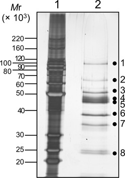
Figure 3.

SDS-10%PAGE analysis of fraction 48 from the MonoS-column chromatography, followed by silver staining. The sizes of markers (lane 1) are shown on the left side of the panel. The proteins in fraction 48 (lane 2) were cut into eight slices, as shown by the dots on the gel, and were subjected to the MS analysis.
