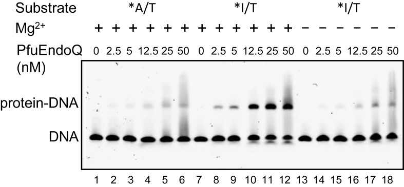
Figure 5.
Electrophoresis mobility shift assay of PfuEndoQ. The dsDNA (5 nM) was incubated with various concentrations of PfuEndoQ (0, 2.5, 5, 12.5, 25 and 50 nM) in the presence (+) (lanes 1–12) or absence (−) (lanes 13–18) of 1-mM MgCl2. *A/T, 5′-Cy5-labeled dsDNA without dI (lanes 1–6); *I/T, 5′-Cy5-labeled dsDNA containing dI (lanes 7–18). The free DNA and the protein–DNA complex were separated by 4% native-PAGE.

