Figure 1.

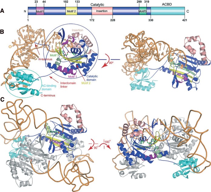
The overall view of the quaternary complexes. (A) The TtHisRS domain architecture. The catalytic, anticodon binding domains (ACBD) as well as the insertion domains are colored blue, cyan and salmon, respectively. The catalytic domain (colored blue) contains three signature motifs 1–3 (colored purple, yellow and pale green, respectively). (B) The side view and the top views of the complex in ribbon rendition. The active site AMP and histidine are shown as sticks while RNA is in orange. (C) The dimeric complex. Only one monomer is colored as described in 1A and the other molecule is colored gray.
