
Figure 1.
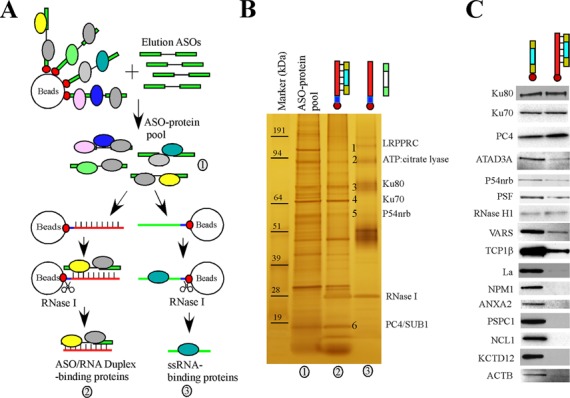
Identification of ASO-binding proteins by affinity selection. (A) Schematic of the affinity selection approaches. The ASO-binding proteins are indicated using colored circles. The biotin tag is indicated by the red dots at the ends of the gapmer ASOs. (B) A representative silver-stained 4–12% SDS-PAGE gel used to resolve affinity-selected proteins. Certain protein bands identified by mass spectrometry are numbered and indicated. Proteins were isolated by competition with ASO (lane 1), ASO/RNA-like duplex (lane 2) or a non-complementary RNA-like 2′-O-methyl oligonucleotide (lane 3). The marker was pre-stained protein Benchmark (Life Technologies). (C) Western analyses of proteins isolated with either ssASO or ASO/RNA-like duplex. Isolated proteins were separated on a 4–12% SDS-PAGE, transferred to a membrane, and probed for indicated proteins by immunoblotting.