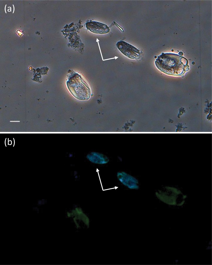
FIG 2.
Bright-field and fluorescence in situ hybridization identification of Charonina ventriculi cells. The images were taken from the same field of fraction 3 of a rumen sample collected from cow C1. (a) Bright-field image. Scale bar, 10 μm. (b) Overlay fluorescence in situ hybridization image of the same field as shown in panel a. Ciliates were simultaneously hybridized with the universal eukaryotic probe EUKb1193 (Cy5) and the Charonina ventriculi-specific probe CHA1350 (Cy3). Color replacement was used to show cells stained with EUKb1193 in green and cells stained with CHA1350 in blue. In the overlay of both images, cyan indicates cells that were stained with both probes. Charonina ventriculi cells are indicated by arrows. Separate images are shown in Fig. S1 in the supplemental material.

