FIG 2.
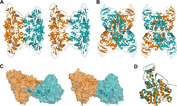
A cruciform quaternary structure is common to MolA and PuhB. (A) Front views of MolA (left) and PuhB (right). (B) Side views of MolA (left) and PuhB (right). (C) C-terminal domain swapping allows extensive salt bridge and hydrogen bond formation across the dimer-dimer interface, which stabilizes the cruciform assembly. MolA has a large C-terminal domain, which enables significant interactions (left). PuhB has a shorter C terminus, which could explain its looser assembly than that of MolA (right). (D) The overall topologies of both enzymes align well. MolA, orange; PuhB, teal.

