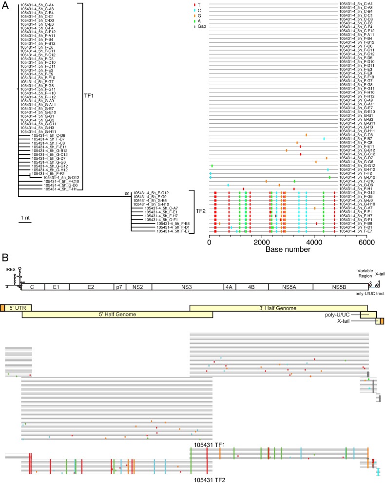
FIG 2 .
Identification of two full-length T/F viral genomes in acutely infected subject 105431. (A) The viral diversity found in subject 105431 is depicted for the 5'-half-genome fragment by a maximum likelihood phylogenetic tree and Highlighter plot. (B) The HCV genome is depicted with gene organization and major RNA secondary structures at the top followed by the amplification strategy for generating full-length HCV genomes in light yellow. Adaptors added to the 5' and 3' termini are depicted in orange. Highlighter plots for each region shown below demonstrate that a high degree of homology is maintained throughout the genomes within the T/F lineages, which are distinguished by large sets of shared polymorphisms.

