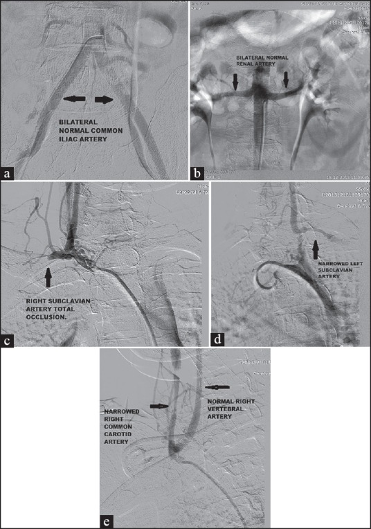
Figure 2.

(a) Digital subtraction peripheral angiography showing normal bilateral common iliac artery. (b) Digital subtraction peripheral angiography showing normal bilateral renal artery. (c) Digital subtraction peripheral angiography showing total occlusion of right subclavian artery.(d) Digital subtraction peripheral angiography showing narrowing of left subclavian artery. (e) Digital subtraction peripheral angiography showing narrowing of right common carotid artery and normal vertebral artery
