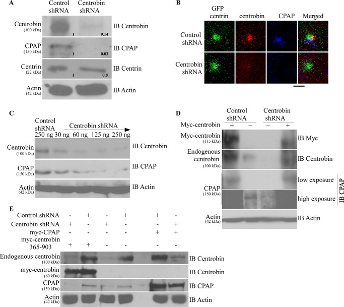
FIGURE 3.
Centrobin depletion results in the degradation of cellular CPAP. A, 293T cells were transfected with control or centrobin shRNA for 72 h after which cells were lysed and probed with the anti-centrobin, -CPAP, -centrin, and -actin antibodies. Relative densitometric values are shown. Individual values were normalized against actin values, and the band intensities of control lane were considered as 1 for calculating relative values. IB, immunoblot. HeLa-GFP-centrin stable cells were transfected as described for A, and centrioles were imaged using the Olympus FV10i confocal microscope. Images are maximum projections of Z stacks. Scale bar: 2 μm. C. 293T cells were transfected with increasing amounts of centrobin shRNA- expressing vector for 72 h, and the lysates were immunoblotted using the indicated antibodies. D, to test the specificity of centrobin shRNA-induced effect, control or centrobin shRNA-transfected 293T cells after 48 h were re-transfected with myc-centrobin-expressing vector. Lysates were probed with antibodies against centrobin, CPAP, and actin. E, same experimental scheme as D, except control or centrobin shRNA cells were re-transfected with myc-centrobin-365–903 or myc-CPAP expressing vectors.

