FIGURE 4.

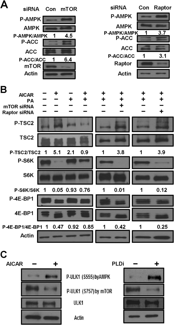
Effects of PLD on AMPK are mediated by mTORC1. A, MDA-MB-231 cells were plated as described for Fig. 3A and transfected with mTOR, Raptor, or scrambled control (Con) siRNA as indicated. After 72 h of transfection, cells were harvested and analyzed for phospho-AMPK (P-AMPK), AMPK, phospho-ACC (P-ACC), ACC, mTOR, Raptor, and actin by Western blot analysis. The relative levels of AMPK and ACC phosphorylation were normalized to total AMPK and total ACC, respectively, and quantified as described for Fig. 1A. B, MDA-MB-231 cells were plated and transfected with mTOR or Raptor siRNA as described for A. After 72 h, cells were treated with AICAR and/or PA as indicated for 45 min. The cells were then harvested, and the levels of phospho-TSC2 (P-TSC2), TSC2, phospho-S6 kinase (P-S6K), S6K, phospho-4E-BP1 (P-4E-BP1), 4E-BP1, and actin were determined as described for A. The relative levels of TSC2, S6K, and 4E-BP1 phosphorylation were normalized to total TSC2, total S6K, and total 4E-BP1, respectively, and quantified as described for Fig. 1A. C, MDA-MB-231 cells were plated as described for Fig. 1B and, where indicated, treated with the PLD inhibitors (PLDi) or AICAR for 1 h. The cells were then harvested, and the levels of phospho-ULK1 (P-ULK1; Ser-555 (AMPK site) and Ser-757 (mTOR site)), ULK1, and actin were determined by Western blotting. The data shown are representative of experiments repeated at least two times.
