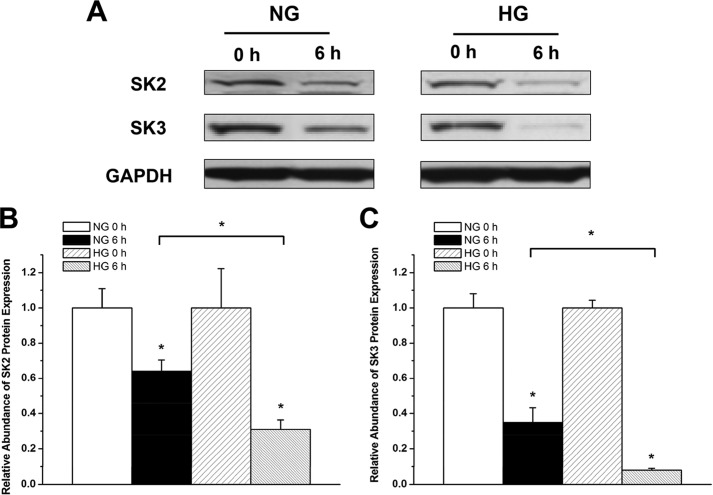
FIGURE 8.
Increased SK2 and SK3 channel protein turnover in HG. A, immunoblots of SK2 and SK3 and GAPDH in HL-1 cells cultured with NG and HG in the presence and absence of cycloheximide (100 μg/ml for 6 h). B, group data showing that SK2 protein turnover was significantly accelerated in HG culture (n = 3). * represents p < 0.05 versus no treatment with cycloheximide and between cycloheximide-treated cells in NG and HG. C, group data showing that SK3 protein turnover was significantly accelerated in HG culture (n = 3). * represents p < 0.05 versus no treatment with cycloheximide and between cycloheximide treated-cells in NG and HG. Error bars represent S.E.

