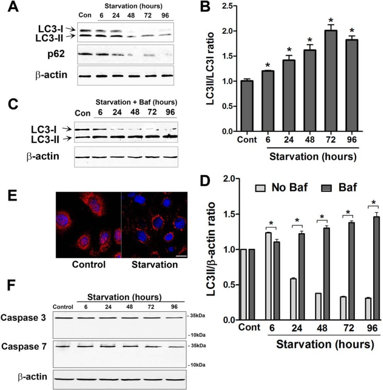
FIGURE 4.
Starvation induces autophagy in Caco-2 cell monolayers. A, Caco-2 cells were incubated in starvation medium for the indicated time and analyzed by Western blotting for LC3B and p62 protein. β-actin is shown as a loading control (Con). B, the ratio of LC3-II/LC3I density was calculated using ImageJ software. The LC3-II/LC3I ratio in the control (Cont) group was set to 1. The Western blot in A and the graph are representative of more than four independent experiments (*, p < 0.01 versus control). C, Caco-2 cells were incubated in starvation medium and bafilomycin A (20 nm) for the indicated time and analyzed by Western blotting for LC3B protein level. D, autophagic flux is represented by the accumulation of LC3II in the presence of bafilomycin A (Baf) (as shown in C) and not in the presence of bafilomycin A (as shown in A) during starvation (*, p < 0.01). E, confocal immunofluorescence for LC3 staining in starved cells showed cytoplasmic punctum formation. Scale bar = 10 μm. Shown is a representation of 48 h of starvation. F, starvation did not induce apoptosis in Caco-2 cells. Caspase-3 and caspase-7 protein expression did not change during starvation. Also, cleaved caspase-3 and -7 were not observed during starvation. Blots are representative of three independent experiments.

