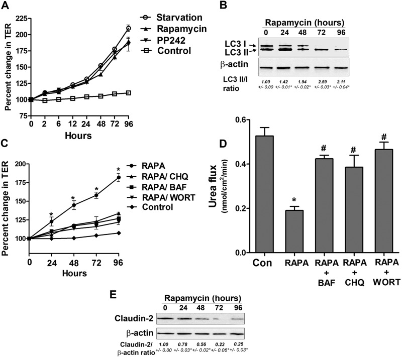
FIGURE 5.
Rapamycin-induced autophagy enhances TJ barrier function. A, incubation of Caco-2 cells in normal medium with the mTOR inhibitors rapamycin and PP242 (500 nm) increased TER compared with cells in starvation medium. B, Caco-2 cells were treated with rapamycin for the indicated time and analyzed by Western blotting for LC3B protein level. β-actin is shown as a loading control. The LC3 II/I densitometry ratio was found to be increased during rapamycin treatment. *, p < 0.01 versus time 0. C, the rapamycin (RAPA)-induced increase in TER was inhibited by the autophagy inhibitors bafilomycin A (BAF, 20 nm), chloroquine (CHQ, 20 μm), and wortmannin (WORT, 200 nm). *, p < 0.01 versus all other groups. D, rapamycin treatment reduced the paracellular flux of urea compared with control cells. The autophagy inhibitors bafilomycin A, chloroquine, and wortmannin attenuated the rapamycin-induced reduction in urea flux. The flux was measured after 96 h of rapamycin treatment. *, p < 0.01 versus control; #, p < 0.01 versus rapamycin. E, rapamycin treatment of Caco-2 cells in normal media led to a reduction in claudin-2 protein level. β-actin is shown as a loading control, and densitometry is represented as the claudin-2:β-actin ratio. *, p < 0.01 versus time 0.

