
Figure 2.
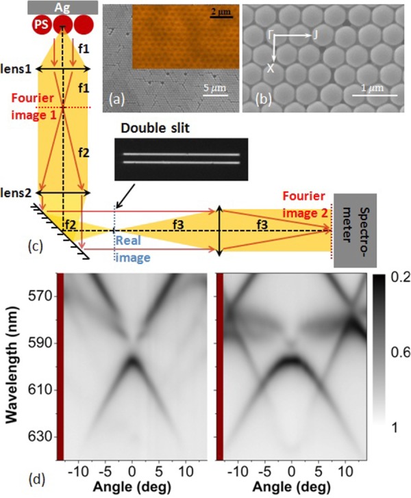
(a, b) The top-view SEM images of the photonic–plasmonic crystal structures in different magnifications. Inset of (a) corresponding to the fluorescence molecules distribution on top of the PS sphere array measured by an oil-immersed confocal fluorescence microscope. (c) The schematic view of the experimental setup, the used double-slit is shown in the inset. (d) Reflection spectra (without a double-slit) of the proposed structure as functions of the wavelength and the incident angle. The light is incident along the Γ–J direction. The left (right) panel is for p- (s-) polarized incident light.