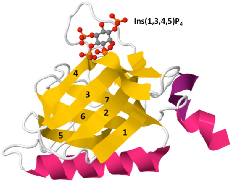
Figure 3.
Crystal structure of Btk PH domain in complex with Ins(1,3,4,5)P4 (PDB ID: 2Z0P). The PH domain is comprised of a β-barrel formed by seven β-strands (yellow, 1–7) capped by an α-helix (pink). The hyper-variable loops of the β-barrel form the binding surface for lipid ligands such as Ins(1,3,4,5)P4 [top, shown by ball-and-stick model: red (oxygen), orange (phosphorus), and gray (carbon)].

