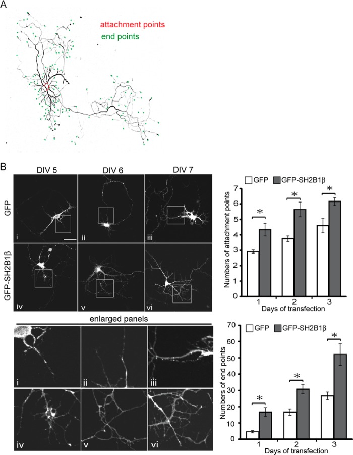
FIGURE 2.
SH2B1β increases branches of neurite in development of hippocampal neurons. A, hippocampal neurons were transiently transfected with GFP-SH2B1β on DIV 3 and images of neuronal morphology were visualized on DIV 7. Attachment points and end points are shown as red and green dots, respectively. B, hippocampal neurons were transiently transfected with either GFP or GFP-SH2B1β on DIV 4, and images of neuronal morphology on days 1–3 after transfection were visualized. Enlarged images of the neurite branches are shown on the bottom panels. Scale bar, 40 μm. A total of 12 hippocampal neurons were counted per condition from three independent experiments. Values are mean ± S.E. from three independent experiments and statistically compared by Student's t test (*, p < 0.05, overexpressing GFP-SH2B1β compared with GFP). The number of attachment points and end points were measured using ImageJ software.

