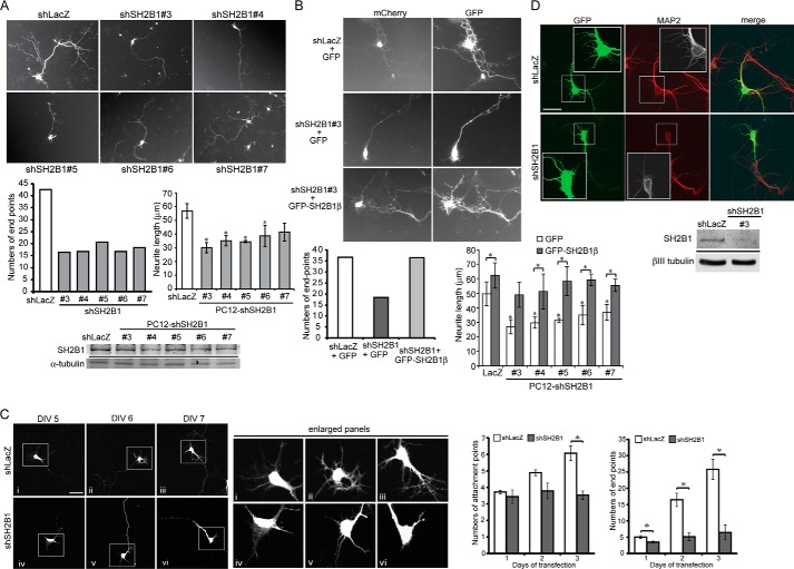
FIGURE 3.
Knockdown of SH2B1 expression reduces neurite outgrowth for hippocampal neurons and PC12 cells. A, hippocampal neurons were transiently transfected with either shLacZ or shSH2B1#3, #4, #5, #6, or #7 together with the pEGFP vector on DIV 4, and live cell images of neuronal morphology were taken. The number of end points was counted from 12 to 18 hippocampal neurons per condition. PC12 cells stably expressing shLacZ or shSH2B1 constructs (shSH2B1#3, #4, #5, #6, and #7) were treated with 100 ng/ml NGF for 1 day. Length of the longest neurite of each cell was calculated from three independent experiments. *, p < 0.05 by paired Student's t test compared with shLacZ control. Lysates from PC12-shLacZ and PC12-shSH2B1 cells were collected and immunoblotted with anti-SH2B1 and anti-α-tubulin antibodies. B, hippocampal neurons were transiently transfected with either shLacZ+mCherry vector or shSH2B1#3+mCherry vector, together with GFP or GFP-SH2B1β on DIV 4, and live cell images were taken. The number of end points was counted from a total of 10–13 hippocampal neurons per condition. PC12-shLacZ or PC12-shSH2B1(#3, #4, #5, #6, and #7) cells were transiently transfected with GFP or GFP-SH2B1β constructs. Transfected cells were then treated with NGF for 1 day, and the length of the longest neurite of each cell was measured from three independent experiments. *, p < 0.05 by paired Student's t test compared with PC12-shLacZ+GFP control or with its respective control. C, hippocampal neurons were transiently transfected with either shLacZ or shSH2B1 together with pEGFP vector on DIV 4, and images of neuronal morphology on days 1–3 after transfection were taken. The number of attachment points or end points on days 1–3 after transfection was measured. Enlarged images of the neurite branches are shown on the right panels. Scale bar, 40 μm. A total of 12 hippocampal neurons were counted per condition from three independent experiments. Values are mean ± S.E. from three independent experiments and statistically compared by Student's t test (*, p < 0.05, shSH2B1 compared with shLacZ). D, hippocampal neurons were transfected with either shLacZ or shSH2B1 together with pEGFP vector on DIV 4, and the images of neuronal morphology of were visualized on DIV 7. Immunofluorescence staining was performed with MAP2 (red), GFP (green), and DAPI (blue) antibodies. Cell lysates from cultured hippocampal neurons transiently transfected with shLacZ or shSH2B1(#3) were collected and analyzed via Western blotting with anti-SH2B1 or anti-βIII tubulin antibody.

