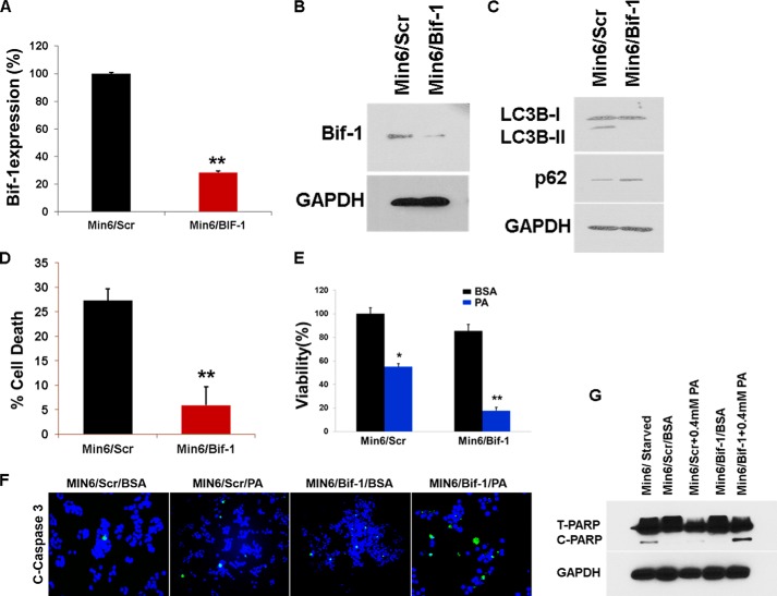
FIGURE 8.
Knockdown of Bif-1 enhances PA-induced cell death. A and B, lentiviral shRNA was used for Bif-1 knockdown. Bif-1 expression was analyzed by RT-PCR (A) and Western blotting (B) after 24 h. **, p < 0.01 versus the level of Bif-1 after infection of scrambled shRNA. C, the MIN6 Bif-1−/− and the scrambled cells were treated with PA (0.4 mm) for 16 h, and the lysates were analyzed by Western blot for SQSTM1/p62 (middle panel) or LC3B and GAPDH (bottom panel). D, the MIN6 Bif-1−/− and the scrambled cells were cultured in HBSS medium for 12 h, and the percentage of cell death was determined by trypan blue exclusion assay (error bars represent S.D. of the mean value from three independent experiments). E, the MIN6 Bif-1−/− and the scrambled cells were treated with PA (0.4 mm) for 16 h. Cell viability was analyzed using Cell Titer-Glo luminescent cell viability assay. *, p < 0.05; **, p < 0.01 versus viability of BSA-treated cells. F, after treating with PA (0.4 mm) for 16 h, immunofluorescence with cleaved caspase 3 antibody was done. G, after treating with PA (0.4 mm) for 16 h, lysates were analyzed by Western blot for cleaved PARP and GAPDH (bottom panel).

