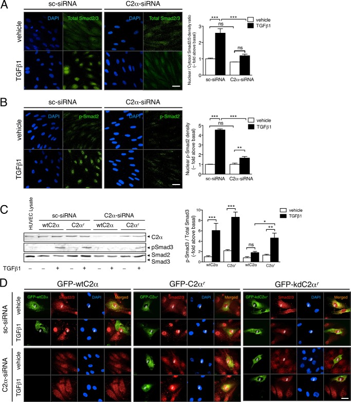
FIGURE 3.
TGFβ1-induced nuclear translocation of Smad2/3 depends on PI3K-C2α in EC. A and B, immunofluorescent staining of Smad2/3 (A) and p-Smad3 (B) in TGFβ1-stimulated HUVECs. The cells were transfected with C2α#1 siRNA or sc-siRNA and stimulated with TGFβ1 (5 ng/ml) for 30 min, followed by anti-Smad2/3 antibody or anti-p-Smad3 antibody staining. Nuclei were stained by DAPI. Left panels, representative confocal images of the stained cells. Right panels, quantified data. The data were obtained from 48 cells per group. Scale bar, 20 μm. C, HUVECs were transfected with either wtC2α or C2α-siRNA-resistant C2α (C2αr) and either C2α#1 siRNA or sc-siRNA. The cells were stimulated with TGFβ1 (5 ng/ml) for 30 min, followed by immunoblot analysis for p-Smad3. Left panel, representative blots. Right panel, quantified data (n = 3). D, HUVECs were transfected with either GFP-wtC2α, GFP-C2αr, or GFP-tagged kinase deficient mutant of C2αr (GFP-kdC2αr) and either C2α-specific siRNA or sc-siRNA. The cells were stimulated with TGFβ1 (5 ng/ml) for 30 min, followed by immunofluorescent staining with anti-Smad2/3. Nuclei were stained by DAPI. Scale bar, 20 μm. #, transfected cells.

