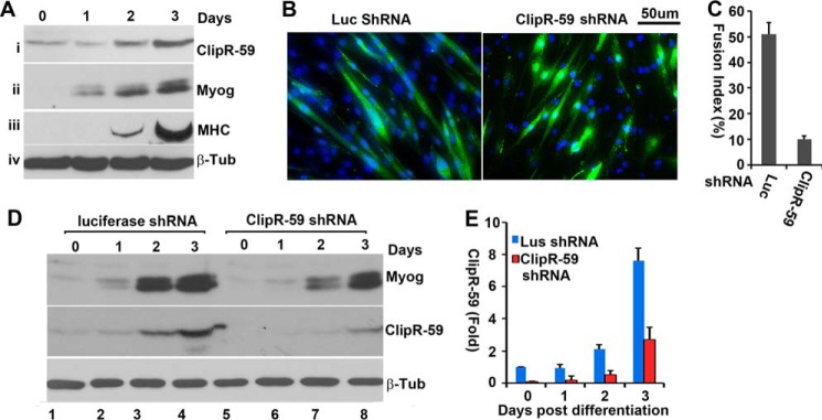
FIGURE 1.

ClipR-59 modulates myoblast fusion. A, ClipR-59 expression during C2C12 muscle differentiation. Total cell lysates from C2C12 cells that had undergone differentiation at the indicated time points were analyzed in Western blot with anti-ClipR-59 (i), anti-myogenin (ii), anti-myosin heavy chain (MHC, iii), and anti-β-tubulin (iv) antibodies, respectively. B, ClipR-59 shRNA expression in C2C12 cells suppressed myoblast fusion. C2C12 cells were transduced with lentiviral vectors that expressed luciferase, and ClipR-59 shRNA, respectively. 48 h post-transduction, cells were switched into differentiation medium (DMEM supplemented with 2% horse serum). 72 h later, the cells were fixed and stained with DAPI. The scale bar represents 50 μm. C, quantification of C2C12 myoblast fusion. The fusion index is the ratio of GFP positive cells that contains more than 3 nuclei and total GFP cells. Bar graphs show mean ± S.D., n = 9 (fields). D, Western blot analysis of total cell lysates collected from the cells in B with anti-myogenin (MyoG), ClipR-59, and β-tubulin antibodies, respectively. E, densitometric analysis of ClipR-59 expression in D. The amount of ClipR-59 in luciferase shRNA expressing C2C12 cells before differentiation was set as 1 after normalized to β-tubulin. The bar graphs show ± S.D. (n = 3). In all cases, p < 0.05.
