FIGURE 3.
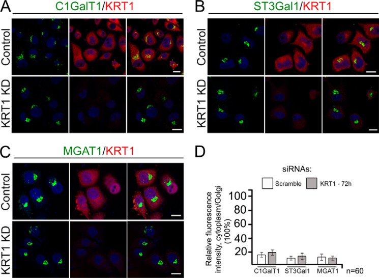
KRT1 was dispensable for Golgi localization of C1GalT1, ST3Gal1, and MGAT1. Shown are confocal immunofluorescence images of C1GalT1 (A), ST3Gal1 (B), MGAT1 (C), and KRT1 in Panc1-bC2GnT-M-c-Myc cells treated with scramble or KRT1 siRNAs for 72 h. Nuclei were counterstained with DAPI (blue). All confocal images were acquired with same imaging parameters; bars, 10 μm. D, quantification of the fluorescence intensity of cytoplasm/Golgi for indicated GTs in cells shown in A–C.

