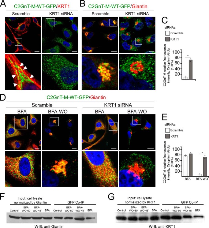
FIGURE 5.
KRT1 was not required for Golgi biogenesis but was critical for Golgi localization of C2GnT-M in HeLa cells. A and B, confocal immunofluorescence images of wild-type C2GnT-M (WT)-GFP colocalization with KRT1 (A) and Giantin (B) in cells treated with scramble or KRT1 siRNAs for 72 h. Arrowheads indicate peri-Golgi colocalization of proteins in control cells. White boxes in the images indicate the area enlarged in the bottom panel that represents merged channels. The results shown are representative of three independent experiments. All confocal images were acquired with same imaging parameters; bars, 10 μm. Nuclei were counterstained with DAPI (blue). C, quantification of relative fluorescence intensity (in a.u.) of C2GnT-M in cytoplasm versus Golgi in cells presented in A and B; *, p < 0.001. D, colocalization of C2GnT-M-WT-GFP and Giantin in cells treated with scramble or KRT1 siRNAs. Next, cells were treated with BFA followed by recovery for 45 min after initiation of BFA WO. E, quantification of relative fluorescence intensity of C2GnT-M in cytoplasm versus Golgi in cells presented in D; *, p < 0.001. F and G, Giantin and KRT1 Western blot (W-B) of complexes pulled down with anti-GFP Ab from the lysates of HeLa cells expressing C2GnT-M (WT)-GFP and treated with BFA followed by WO for 45 and 60 min.

