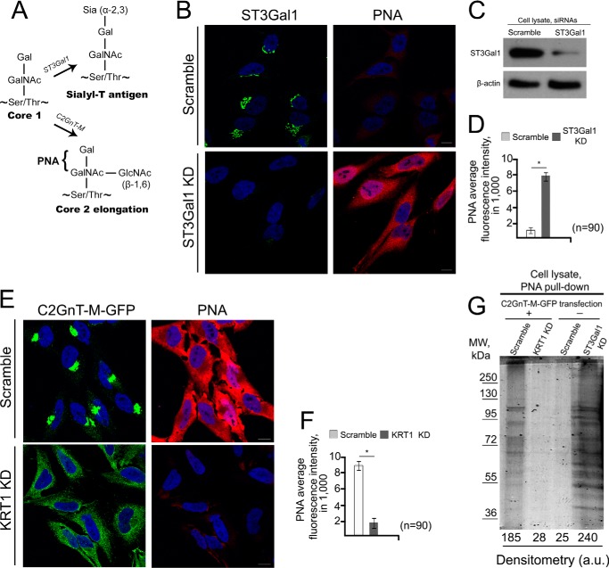
FIGURE 6.
Loss of C2GnT-M Golgi localization was accompanied by the production of sialyl-T antigen. A, schema of selective mucin core 1 and core 2-associated O-glycans in mammalian cells. The core 1 structure may be extended by core 2 as catalyzed by C2GnT-M or alternatively shortened by formation of Siaα2–3Galβ1–3GalNAc (Sialyl-T) antigen as catalyzed by ST3Gal1. The PNA lectin binds non-sialylated core 1 O-glycans, Gal-(±GlcNAc)GalNAc. B, confocal immunofluorescence images of PNA lectin-staining in HeLa cells treated with scramble or ST3Gal1 siRNAs. C, ST3Gal1 Western blot of the lysate of HeLa cells treated with scramble or ST3Gal1 siRNAs for 72 h; β-actin was a loading control. D, quantification of PNA-specific average integrated fluorescence (in a.u.) in the cells shown in B; *, p < 0.001. E, confocal immunofluorescence images of C2GnT-M-WT-GFP and PNA lectin-staining in HeLa cells expressing C2GnT-M-WT-GFP and treated with scramble or KRT1 siRNAs. All confocal images were acquired with the same imaging parameters; bars, 10 μm. Nuclei were counterstained with DAPI (blue). F, quantification of PNA-specific average integrated fluorescence (in a.u.) in the cells shown in E; *, p < 0.001. G, Coomassie Blue staining of the 8% SDS-PAGE for separating the glycoproteins pulled down with biotinylated PNA and then streptavidin magnet beads from the lysates of HeLa cells transiently expressing C2GnT-M-GFP and then treated with scramble or KRT1 siRNAs or wild-type HeLa cells treated with scramble or ST3Gal1 siRNAs.

