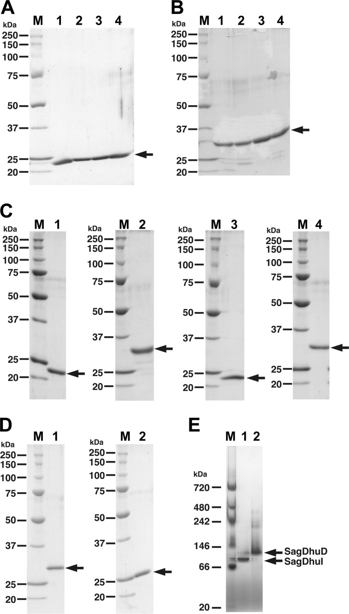
FIGURE 3.
Electrophoretic profile of purified enzymes. A, SDS-PAGE profile of gbs1892 (SagDhuI). Lane 1, wild-type SagDhuI; lane 2, SagDhuI C72A; lane 3, SagDhuI T74A; lane 4, SagDhuI N106A. B, SDS-PAGE profile of gbs1891 (SagDhuD). Lane 1, wild-type SagDhuD; lane 2, SagDhuD S150A; lane 3, SagDhuD Y163F; lane 4, SagDhuD K167A. C, SDS-PAGE profile of spr0289 (SpnDhuI, lane 1), spr0290 (SpnDhuD, lane 2), SPy0637 (SpyDhuI, lane 3), and SPy0636 (SpyDhuD, lane 4). D, SDS-PAGE profile of EcoKduI (lane 1) and PcaKduD (lane 2). Lane M in A–D, molecular weight standards (from top): recombinant polypeptides with molecular weights of 250,000, 150,000, 100,000, 75,000, 50,000, 37,000, 25,000, and 20,000. E, blue native PAGE. Lane 1, gbs1892 (SagDhuI); lane 2, gbs1891 (SagDhuD). Lane M, molecular weight standards (from top): polypeptides with molecular weights of 720,000, 480,000, 242,000, 146,000, 66,000, and 20,000.

