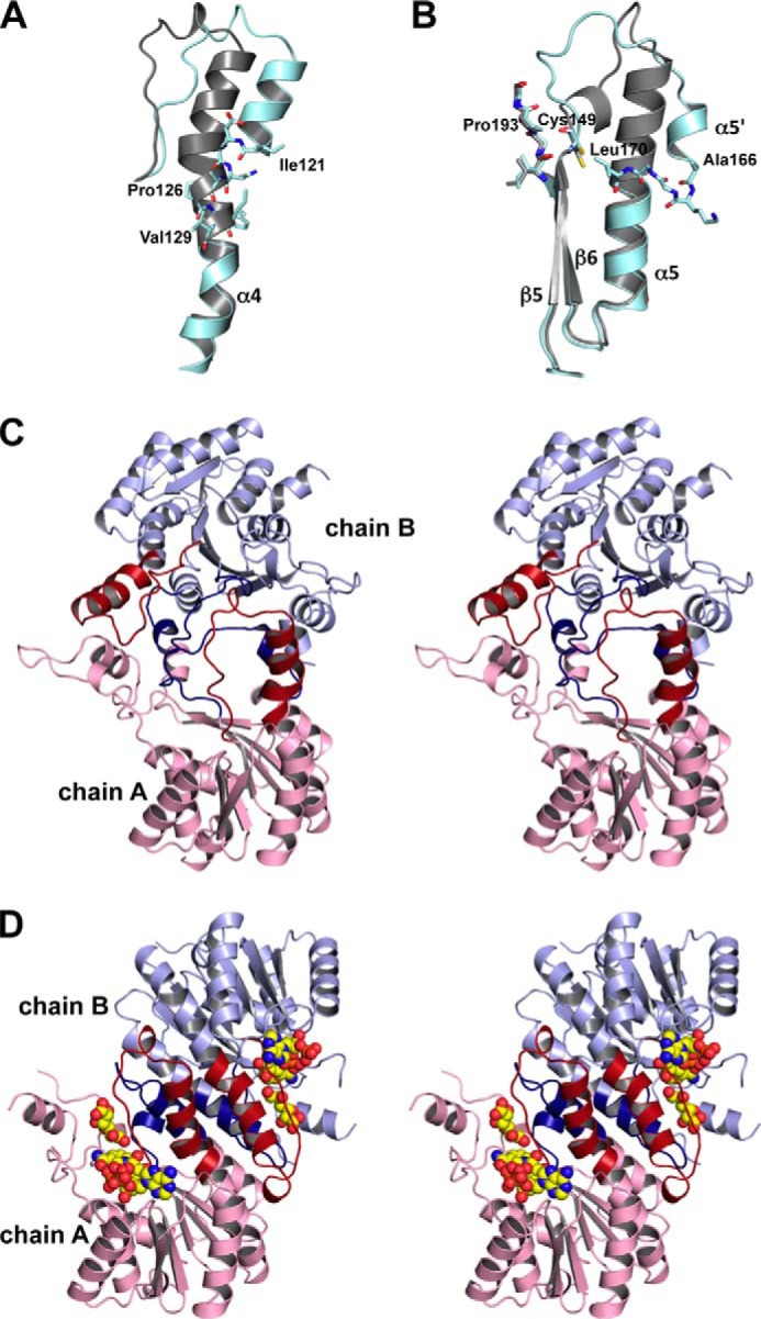
FIGURE 8.

Structural comparison. A, the bent helix 4 in SagDhuD. Cyan, SagDhuD; gray, Ga5DH (PDB code 3CXR). The residues Ile121 and Val129 are indicated by stick models. B, part of helix 5 of SagDhuD (cyan) is loosened distinct from the long helix 5 of Ga5DH (gray). The residues Cys149, Ala166–Leu170, and Ile191–Gly194 are indicated by stick models. C, interaction between two monomeric subunits (chains A and B) (stereo diagram). The bent and shortened helices of SagDhuD are colored red and blue, respectively. D, subunit interaction of substrate/cofactor-bound Ga5DH (PDB code 3O03) (stereo diagram). Helices 4 and 5 are colored in red and blue, respectively. Substrate and cofactor are indicated by sphere models. The orientation of chain A is identical to that of chain A in C.
