FIGURE 1.
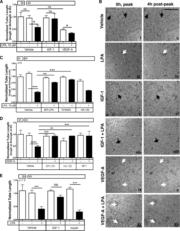
IGF-1 prevented regression of endothelial tubes. A, IGF-1 prevented LPA-stimulated regression of tubes. Endothelial tubes were generated by plating primary HMRECs between two layers of collagen and overlaying the collagen sandwich with serum- and VEGF-containing medium (see “Experimental Procedures”). Tubes formed and stabilized within 16 h under these conditions. The tubes were photographed (0 h, “peak”), serum free medium containing the indicated agents was added, and then the tubes were rephotographed 4 h later (4 h post-peak). For the phase-contrast images acquired at 0 and 4 h, the tubes were traced using the paintbrush function of National Institutes of Health ImageJ. Normalized tube length was calculated by dividing the total pixel units at 4 h (end point) with the total pixel units at 0 h. Data represent the mean ± S.E. of four experiments. Each independent experiment was from quadruplicate wells. *, p < 0.05; ns, not significant (one-way ANOVA). B, representative phase contrast images of HMREC tubes before (0 h, peak) and after (4 h post-peak) addition of the indicated treatments. Black arrows point to representative enduring tubes, and white arrows denote examples of tubes that regressed. Scale bar = 250 μm. C, LPA-mediated regression was blocked with LPA receptor antagonists. Tubes were organized as in A, and regression was monitored in the presence of the indicated agents. BrP-LPA (20 μm) and Ki16425 (20 μm) are LPA receptor antagonists, whereas HA-130 (10 μm) is an ATX inhibitor (23). D, IGF-1 prevented VEGF-A-mediated regression. After generating tubes as described in A, VEGF-mediated regression was assayed in the presence of the indicated agents: VEGF-A (2.5 ng/ml), BrP-LPA (20 μm), HA-130 (10 μm), and IGF-1 (100 ng/ml). E, insulin failed to oppose LPA-stimulated regression. Tubes were organized as in A, and the impact of IGF-1 (100 ng/ml) and insulin (100 ng/ml) to prevent LPA-mediated regression was compared. *, p < 0.05; **, p < 0.01; ***, p < 0.001; ****, p < 0.0001.

