FIGURE 3.
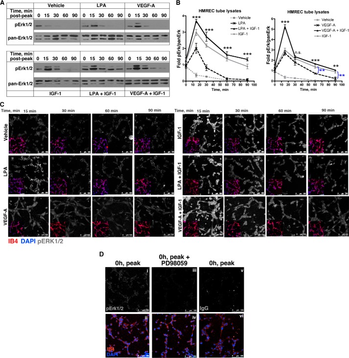
IGF-1-mediated stabilization was associated with enduring activation of Erk. A, HMREC tubes were generated as described in Fig. 1A, except that these experiments were performed in 24-well plates. At the indicated time points, tube lysates were extracted and subjected to Western blot analysis. Five wells were pooled for each lane/time point. B, the data from three to six independent Western blot experiments were quantified and expressed as a ratio of pErk:panErk. These values were normalized to the average of the unstimulated samples (the 0 time point in A), and the mean ± S.E. was plotted. Asterisks indicate paired comparisons between LPA or VEGF alone versus LPA + IGF-1 or VEGF + IGF-1. **, p < 0.01; ***, p < 0.001; ns, not significant (Student's t test). C, IGF-1 induced enduring activation of Erk. The lower left quadrant shows an inset of the same tubes that were costained with isolectin B4 (which decorates endothelial cells, red) and DAPI (nuclei, blue). The remainder of each panel is the same magnification as the lower left quadrant and shows the staining with an anti-pErk1/2 antibody (shown in monochrome). Images are representative of three independent experiments performed in duplicate wells (3 random fields/well). Scale bar = 100 μm. D, controls to assess the specificity of anti-pErk1/2 immunofluorescent signal. When tubes had formed (0 h, peak, as in Fig. 1B), they were stained with the anti-pErk1/2 antibody (i) or a non-immune isotype IgG (vi). Alternatively, tubes were pretreated with an Erk pathway inhibitor (PD98059, 10 μm) and stained with the anti-pErk1/2 antibody. The bottom row (ii, iv, and vi) shows and IB4 and DAPI-stained channels of the top row.

