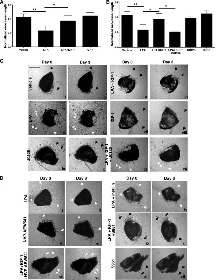
FIGURE 5.
IGF-1 prevented LPA-dependent regression of retinal neovessels, and this phenomenon was dependent on Erk activity. A, IGF-1 antagonized LPA-mediated regression of retinal neovessels. Murine retinal explants were placed in a collagen sandwich and overlaid with serum- and VEGF-containing medium. Neovessels sprouted within 7–10 days and, thereafter, increased in number, length, and caliber through days 18–21. Neovessels that showed no significant outgrowth within a 48-h window were considered stable. Stable neovessels were photographed (day 0), endothelial basal medium with 10% serum (vehicle) alone or in combination with the indicated treatments was added, and the neovessels were rephotographed 3 days later (day 3). The treatments included LPA (10 μm), IGF-1 (100 ng/ml), the MEK inhibitor U0126 (10 μm), insulin (100 ng/ml), the IGF-1R tyrosine kinase inhibitor NVP-AEW541 (10 μm), and the insulin receptor antagonist S961 (100 nm). Treatments were replenished every 12 h within this 3-day window. For the phase-contrast images acquired at days 0 and 3, the neovessels were traced using the paintbrush function of National Institutes of Health ImageJ. Normalized neovessel length was calculated by dividing the total pixel units at day 3 (end point) with the total pixel units at day 0. Data represent the mean ± S.E. of three mice, and each independent experiment was performed in duplicate (left eye and right eye). *, p < 0.05; **, p < 0.01 (one-way ANOVA). B, blocking the Erk pathway overcame the ability of IGF-1 to prevent LPA-mediated regression of neovessels. Experiments in A were repeated in a parallel assay wherein the retinal explants were treated with the MEK inhibitor U0126 at a dose of 10 μm. Data represent the mean ± S.E. of three mice, and each independent experiment was performed in duplicate (left eye and right eye). *, p < 0.05; **, p < 0.01 (one-way ANOVA). C and D, representative phase-contrast images of retinal explants at days 0 and 3. Black arrows point to representative neovessels that did not regress, and white arrows denote examples of neovessels that did regress. Scale bar = 250 μm.

