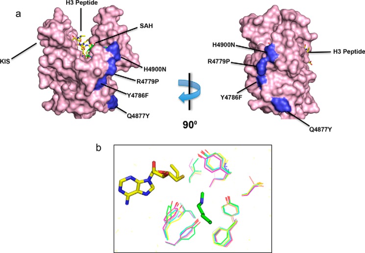
FIGURE 7.
a, surface representation of the crystal structure of the MLL1 SET domain bound to histone H3 peptide (yellow) and S-adenosyl-l-homocysteine (SAH) (green) (Protein Data Bank code 2W5Z) (67). The position of the four gain-of-function mutants are highlighted in blue and noted with their MLL3 numbering. The location of the previously identified Kabuki interaction surface (KIS) is noted. b, homology modeling of the conserved active site residues in the human SET1 family predicts that they adopt similar three-dimensional positions. Models were generated in Modeler (68) using the MLL1 structure (PDB code 2W5Z) as a template.

