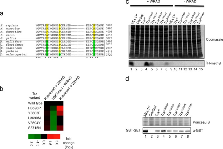
FIGURE 8.
Gain-of-function positions identified in MLL3 enhance WRAD-dependent dimethylation by the complex assembled with Drosophila Trx. a, representative alignment of MLL1 orthologs reveals amino acid positions that show a split between invertebrates (boxed) and vertebrates. b, heat map of methyltransferase activities of wild type and mutant Trx constructs. The data represent the log2-fold change (mutant/wild type) in activity of Trx constructs with the indicated peptide. c, representative gel of gain-of-function Trx mutants. The upper panel shows a Coomassie Blue-stained SDS-polyacrylamide gel, and the lower panel shows [3H]methyl incorporation after 24 h of fluorography. d, assessment of the expression level of mutant Trx constructs. Lysate samples from mutant and wild type constructs were separated by SDS-PAGE, transferred to PVDF membranes, and blotted with an α-GST antibody. The upper panel shows a Ponceau S-stained PVDF membrane, and the lower panel shows the Western blot. The control represents an untransformed E. coli lysate that was induced with 0.75 mm IPTG.

