FIGURE 3.
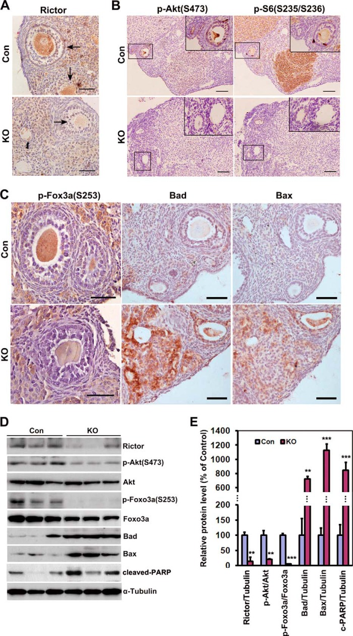
Elimination of Rictor in oocyte replicates the toxicological effects of VCD on Rictor/mTORC2 signaling. A, immunohistochemical analysis showed successful deletion of Rictor in oocytes of cKO mice. No Rictor expression was observed in oocytes of cKO mice. Con, control. B, reduced Ser-473-phosphorylated Akt and Ser-235/Ser-236-phosphorylated S6 in ovaries of cKO mice, particularly in corpus luteum. The inset represents an enlargement of the area in the box. C, decreased level of Ser-253-phosphorylated Foxo3a simultaneously observed with enhancement of Bad, Bax, and cleaved PARP in oocytes of cKO mice. D, Western blot analysis for comparison of Rictor/mTORC2 signaling in the ovaries of control and cKO mice. Protein samples were extracted from the whole ovary. Three individual mice of each group are shown. E, quantification of the results of D. Ovaries were from 6-month old control and cKO mice. Bars indicate mean ± S.E. **, p < 0.01, ***, p < 0.001. Scale bar = 50 μm for A and C, 200 μm for B.

