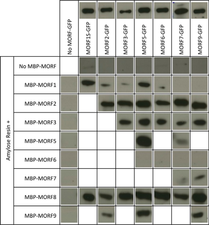
FIGURE 4.

Pulldown probings of MORF-MORF protein interactions. Shown are the respective MORF-GFP GFP antibody chemiluminescence signals in the respective gel lanes pulled down with the respective MORF-MBP proteins bound to an amylose resin. MORF8 interacts with most other MORF proteins, whereas e.g. MORF7 binds only a few other MORF proteins. Without any MBP-MORF protein, no GFP-labeled MORF is retained by the amylose resin, and without a GFP-tagged MORF, no GFP antibody signal is detected. Above the respective MORF-GFP lanes, loading controls of 1 μg of the respective protein preparation are shown. Protein concentrations were determined by Bradford assays, and differences in the actual amounts of the respective GFP protein are due to variations of the impurities carried over from the overexpression in E. coli. For the pulldown assays, 10 μg of the respective GFP protein preparation was used. MORF1S-GFP is shortened in the C-terminal region outside of the MORF domain. Details are discussed under “Results” and in the legend for Fig. 6.
