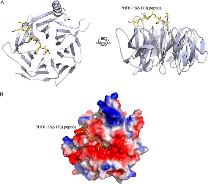
FIGURE 2.
Crystal structure of the PHF6 peptide bound to RBBP4. A, two orthogonal views of RBBP4 bound to the PHF6 peptide, with the N and C termini of RBBP4 labeled. The PHF6 peptide is shown in yellow. B, electrostatic surface of RBBP4 showing the PHF6 peptide-binding pocket. The PHF6 peptide is shown as in A.

