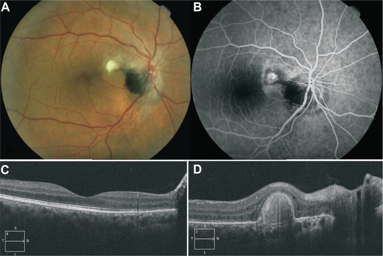
Figure 2.
Post treatment fundus color image, fluorescein angiography and optical coherence tomography findings.
Notes: (A) Color image of the right eye fundus of the same patient 3 years after treatment showing complete resolution of subretinal fluid, lipid deposits and hemorrhage, and a fibrotic scar at the level of the choroidal neovascular complex; an apparent subretinal extension from the superotemporal margin of the tumor extends as a tongue toward the choroidal neovascular scar; inframacular and infrapapillar subretinal punctuate pigment dispersion is also observed, and adjacent choroidal component melanocytoma is now visible at the inferior margin of the optic disk. (B) Venous phase of fluorescein angiogram showing impregnation of choroidal neovascular scar and no capillary telangiectasia and juxtapapillar and optic disk leakage. (C) Spectral domain optical coherence tomography scan passing through the macula showing complete resolution of subretinal fluid and macular edema. (D) Spectral domain optical coherence tomography scan passing through choroidal neovascular scar.

