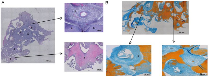
Figure 5.

Haematoxylin-Eosin staining (5A) showing the presence of a bone substitute (S) well integrated in the host bone tissue (B); Trichrome Mallory staining (5B) showing the presence of newly formed bone tissue indicated by orange staining (B) and collagen fibrils (F) represented in blue.
