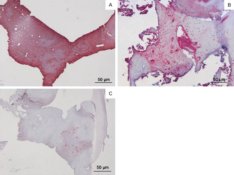
Figure 6.

Immunohistochemical analysis for type I collagen (A) showing a strong positivity at cellular and extracellular matrix level; immunohistochemical analysis for osteocalcin (B) showing a strong positivity at cellular level in almost the entire thickness of the bone areas within biopsy; immunohistochemical analysis for alkaline phosphatase (C) showing a slight positivity at cellular level.
