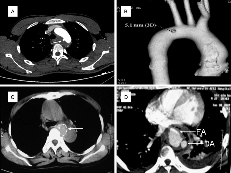
Figure 3.

MDCT subtypes of AEFs Grade IV injuries. A, B: Pseudoaneurysm and AEF, with a diverticulum (white arrow) on the medial wall of the aortic arch, surrounded with inflammatory tissue. C: Fibrous encapsulation and AEF, with a swollen soft-tissue mass (white arrow) between the esophagus and descending aortic wall, whose boundary on the right side is unclear. D: Mediastinal abscess and AEF in the plane of the inferior pulmonary vein. A contrast-enhanced scan shows the fistula on the descending aortic (DA) wall, and the contrast agent flows into the false aneurysm (FA). Inflammatory and necrotic tissue is seen around the esophagus and the descending aorta, with gas formation and left pleural effusion.
