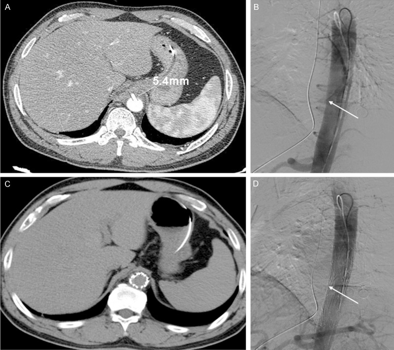
Figure 4.

A: CT angiography images. AEF with inflammatory exudate surrounding the descending aorta (diameter 5.4 mm). B: An aortic pseudoaneurysm at the anterior wall of the descending aorta at the lower edge of thoracic vertebrae 10 (arrow). C, D: Aortic endovascular stent graft implantation to close the AEF (arrow).
