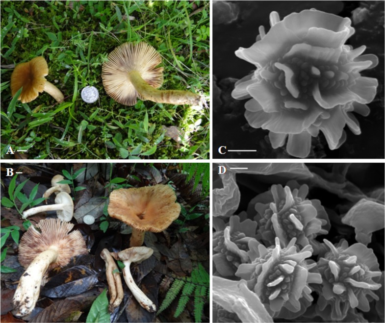
Figure 1. Fresh basidiomata and basidiospore ornamentation of Russula senecis.

(A–B). Basidiomata. (C–D). SEM microphotograph of basidiospores. Bars (A–B): 10 mm; (C–D): 2 µm. Photographer for (A) and (B): Arun Kumar Dutta

(A–B). Basidiomata. (C–D). SEM microphotograph of basidiospores. Bars (A–B): 10 mm; (C–D): 2 µm. Photographer for (A) and (B): Arun Kumar Dutta