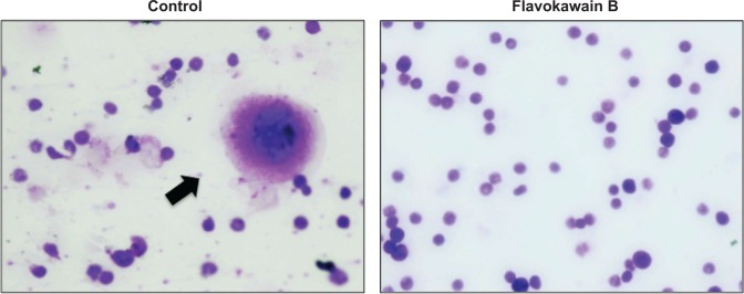
Figure 9.

Bone marrow cells stained with Giemsa viewed under a phase contrast microscope.
Notes: Black arrow indicates the presence of abnormal cells within the cell population. Abnormal cells are identified by the morphology difference. Magnification: 40×.
