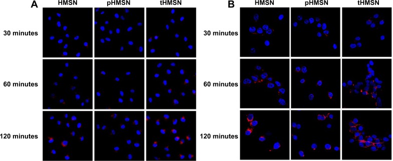
Figure 8.
Confocal laser scanning microscopic images of MDA-MB-231 cells (A) or human umbilical vein endothelial cells (B) incubated with HMSN, pHMSN, or tHMSN loaded with the same amount of doxorubicin for different periods of time. Blue indicates Hoechst 33342. Red indicates doxorubicin.
Abbreviations: HMSN, hollow mesoporous silica nanoparticles; tHMSN, tLyp-1 and polyethylene glycol co-modified HMSN; pHMSN, polyethylene glycol-modified HMSN.

