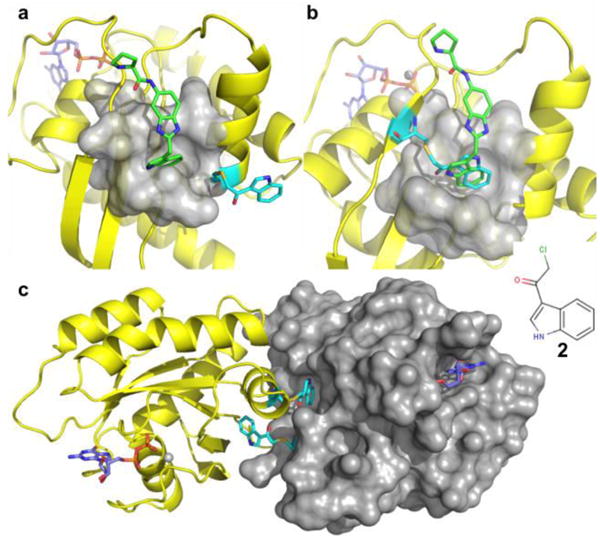
Fig. 2.

Crystal structures of various K-Ras mutants covalently attached to compound 2 (cyan). a) In mutant T74C, the covalently attached compound 2 is pointing towards the solvent. b) In the mutant S39C, the linked compound is sitting inside of the primary pocket, but the NH group of the indole is pointing in the opposite direction compared to compound 1 (green). c) The modified Q70C mutant crystallized as a dimer with the indole portion of the compound occupying the pocket of a neighboring molecule. (PDB code 4PZY) None of these modifications blocks the probe compound 1 from binding to the first-site
