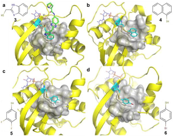
Fig. 3.

Ribbon and molecular surface representations of the crystal structures of GDP-bound K-Ras S39C that was reacted with thiol-reactive compounds (cyan) a) 3, (PDB 4PZZ) b) 4, (PDB 4Q01) c) 5, (PDB 4Q02) and d) 6. (PDB 4Q03) All these compounds completely block the pocket and prevent the probe compound from interacting with the protein. Among them, compound 3 perfectly overlays with the probe compound 1 (green)
